

VASCULAR STUDIES – ULTRASOUND AND DOPPLER (DUPLEX)
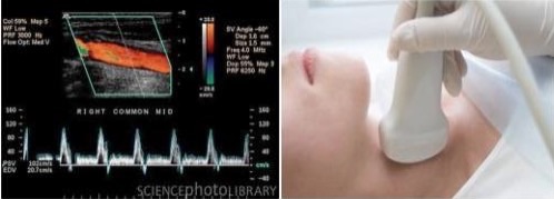
Ultrasound provides 2-dimensional images of the blood vessels and doppler assesses the velocities of blood flow. Color flow imaging is basically doppler data represented in color on a 2-dimensional real-time image. Vital information about the severity of the blockages and composition of the plaques can be obtained. Therefore, it is not surprising that duplex ultrasound studies have become the technique of choice to detect, quantify and follow the progression of vascular disease in different areas:
Carotid arteries: neck arteries that supply blood flow to the brain, (blockages may result in strokes),
Peripheral (leg) arteries: blockages may cause exertional leg pain (intermittent claudication),
Renal arteries: arteries that supply blood to the kidneys, (blockages may result in severe hypertension and renal failure) and,
Aorta: the gradual dilatation or “ballooning” of an artery is called aneurysm, the most common of which is the aneurysm of the abdominal aorta. Growth rate of the aneurysm varies but a mean rate of enlargement of 0.4 cm/yr have been reported. Yearly examinations are necessary to measure the aneurysm size. Surgical treatment is necessary in large aneurysms (greater than 4.5-5 cm in diameter) to prevent rupture and internal bleeding, sometimes fatal. Also first-degree relatives of patients with abdominal aneurysms should probably be screened.